xENi
Experimental Electrophysiology and Neuroimaging
The research focus of the xENi Lab is in systems neuroscience. We are interested in the neuronal coding of sensorimotor function in health and disease with a particular focus on the functional and structural reorganization and non-invasive brain stimulation after stroke. The lab holds a multimodal methodological expertise covering advanced electrophysiology techniques (M/EEG), functional and structure neuroimaging (MRI), non-invasive brain stimulation (tDCS, tACS and TMS), behavioural testing and kinematics as well as data-science and machine-learning methods.
Oscillatory network changes after stroke
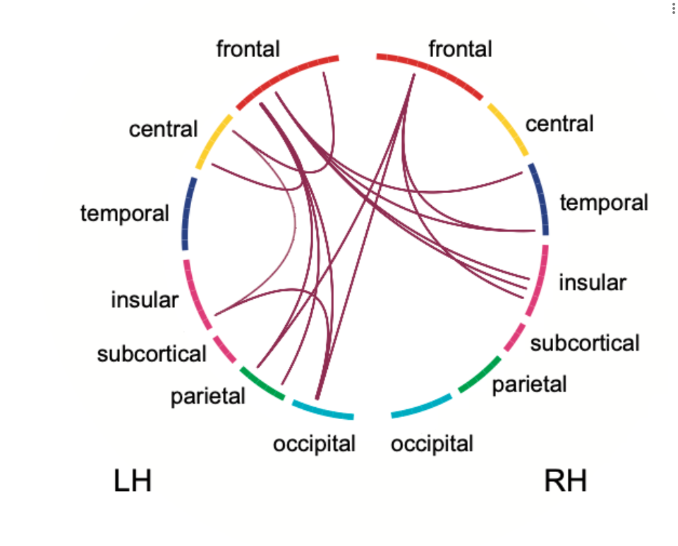
Neural oscillations are considered to play a major role in the information transfer in the brain. Information is transferred not only by fluctuation in the local neuronal population, but by communication through neuronal coherence over larger distances. We investigate oscillatory network changes in cerebrovascular disease, aiming to understand the underlying neurophysiology of the impact of lesions on network changes and recovery and use network features as a biomarker to predict clinical function.
This project is funded by the Hertie Network of Exellence.
Related Publications:
Quandt, F., Fischer, F., Schroder, J., Heinze, M., Kessner, S. S., Malherbe, C., Schulz, R., Cheng, B., Fiehler, J., Gerloff, C., & Thomalla, G. (2020). Normalization of reduced functional connectivity after revascularization of asymptomatic carotid stenosis. J Cereb Blood Flow Metab, 40(9), 1838-1848. https://doi.org/10.1177/0271678X19874338
Quandt, F., Fischer, F., Schroder, J., Heinze, M., Lettow, I., Frey, B. M., Kessner, S. S., Schulz, M., Higgen, F. L., Cheng, B., Gerloff, C., & Thomalla, G. (2020). Higher white matter hyperintensity lesion load is associated with reduced long-range functional connectivity. Brain Commun, 2(2), fcaa111. https://doi.org/10.1093/braincomms/fcaa111
Oscillatory coding of motor control

Dexterous finger movements are a hallmark of human motor control, and its movements are associated with changes in oscillatory brain activity. We investigate cortical oscillations related to motor control using EEG and MEG and relate the neuronal code to behavioral impairment and recovery after stroke. We aim to provide further insight into the role of oscillatory activity in motor control and its mechanisms underlying motor recovery after stroke to identify potential target for noninvasive brain stimulation to improve motor rehabilitation. In a current project we focus on the role of high gamma oscillations in motor control after stroke. The project is conducted in collaboration Dr. Bettina Schwab and funded by internal funds of the UKE and the SFB-936.
Related publications:
Durschmid, S., Quandt, F., Kramer, U. M., Hinrichs, H., Heinze, H. J., Schulz, R., Pannek, H., Chang, E. F., & Knight, R. T. (2014). Oscillatory dynamics track motor performance improvement in human cortex. PLoS One, 9(2), e89576. https://doi.org/10.1371/journal.pone.0089576
Quandt, F., Bonstrup, M., Schulz, R., Timmermann, J. E., Mund, M., Wessel, M. J., & Hummel, F. C. (2019). The functional role of beta-oscillations in the supplementary motor area during reaching and grasping after stroke: A question of structural damage to the corticospinal tract. Hum Brain Mapp, 40(10), 3091-3101. https://doi.org/10.1002/hbm.24582
Quandt, F., Bonstrup, M., Schulz, R., Timmermann, J. E., Zimerman, M., Nolte, G., & Hummel, F. C. (2016). Spectral Variability in the Aged Brain during Fine Motor Control. Front Aging Neurosci, 8, 305. https://doi.org/10.3389/fnagi.2016.00305
Quandt, F., Reichert, C., Hinrichs, H., Heinze, H. J., Knight, R. T., & Rieger, J. W. (2012). Single trial discrimination of individual finger movements on one hand: a combined MEG and EEG study. Neuroimage, 59(4), 3316-3324. https://doi.org/10.1016/j.neuroimage.2011.11.053
Schulz, R., Bonstrup, M., Guder, S., Liu, J., Frey, B., Quandt, F., Krawinkel, L. A., Cheng, B., Thomalla, G., & Gerloff, C. (2021). Corticospinal Tract Microstructure Correlates With Beta Oscillatory Activity in the Primary Motor Cortex After Stroke. Stroke, 52(12), 3839-3847. https://doi.org/10.1161/STROKEAHA.121.034344
Oscillatory encoding of kinematic features after stroke
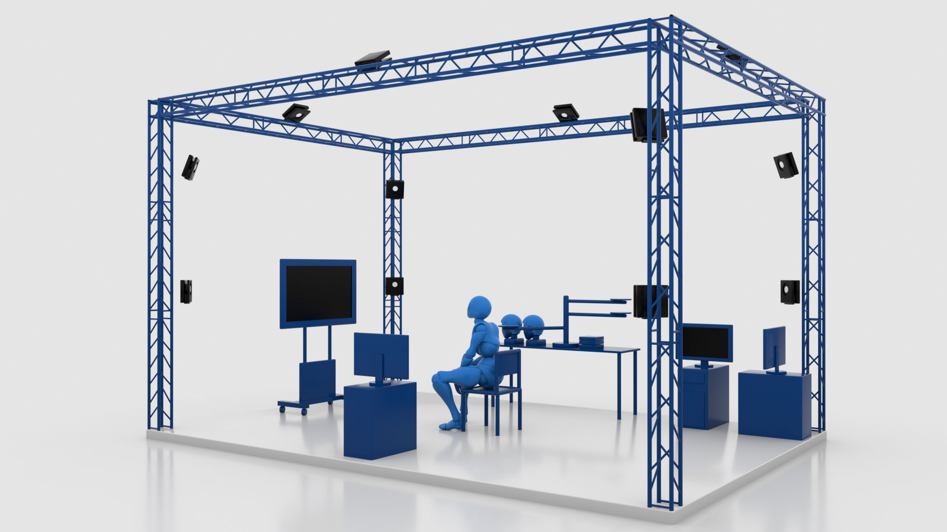
This project has the principal goal of advancing our understanding of how neuronal networks in the human brain reorganize after a stroke and how this affects complex motor behavior relevant to everyday life. We try to gain new insights into motor-related cognitive processes, and to expand our network models through a technically innovative analysis of the spinal cord’s activity. By using an advanced system of infrared 3D cameras while measuring EEG signals we are able to study kinematic parameters, such as “smoothness” and “directness” of the movements, and to correlate them with the brain activity of the participants. Additionally, we are able to study the activation in predefined cortico-spinal networks with special fMRI-sequences. In addition to gaining fundamental scientific knowledge, we also study the neuroplasticity-enhancing effects of targeted neuromodulation of neural networks.
This project was part of the Collaborative Research Center 936 – Multi-Site Communication in the Brain (CRC 936/SFB 936) and funded by the Deutsche Forschungsgesellschaft (DFG). DFG Project number, 178316478
Multisensory integration in health and disease

The overall goal of the project is to examine the neural dynamics of multisensory integration in healthy older adults and clinical cohorts including stroke. We analyze neural signals associated with visuo-tactile integration and learning using M/EEG and functional MRI. Specifically, we are interested in the interrelation between the structural and functional changes in multisensory brain networks. Therefore we simultaneously record EEG and fMRI data. Furthermore, we employ non-invasive brain stimulation to modulate related brain areas in attempt to enhance performance and aim to build computational models. Our investigations focus specifically on oscillatory neural activity and large-scale dynamic coupling between brain regions. Sensory feedback and virtual reality (VR) settings will be used to to modulate and, ideally, improve performance in crossmodal integration tasks.
This project ist part of the Transregional Collaborative Research Centre – Crossmodal Learning: Adaptivity, Prediction and Interaction (TRR 169) and funded by the German Research Foundation (DFG) and the National Natural Science Foundation of China (NSFC).
Structural brain reserve

The concept of brain reserve capacity positively influencing the process of recovery after stroke has been continuously developed in
recent years. Global measures of brain health such as brain volumes or burden of small vessel disease have been linked with a favourable outcome. Our lab is interested in the importance of MRI-based structural reserve estimates for recovery after stroke, i.e., the pre-stroke state of important brain regions such as contralesional motor cortices, the cerebellum, and basal ganglia.
Related Publications:
Sadeghihassanabadi F, Frey BM, Backhaus W, Choe CU, Zittel S, Schon G, Bonstrup M, Cheng B, Thomalla G, Gerloff C, et al. Structural cerebellar reserve positively influences outcome after severe stroke. Brain Commun. 2022;4:fcac203. doi: 10.1093/braincomms/fcac203
Rojas Albert A, Backhaus W, Graterol Perez JA, Braabeta H, Schon G, Choe CU, Feldheim J, Bonstrup M, Cheng B, Thomalla G, et al. Cortical thickness of contralesional cortices positively relates to future outcome after severe stroke. Cereb Cortex. 2022;32:5622-5627. doi: 10.1093/cercor/bhac040
Asmussen L, Frey BM, Frontzkowski LK, Wrobel PP, Grigutsch LS, Choe CU, Bonstrup M, Cheng B, Thomalla G, Quandt F, Gerloff C, Schulz R. Dopaminergic mesolimbic structural reserve is positively linked to better outcome after severe stroke. Brain Commun. 2024;6:fcae122. doi: 10.1093/braincomms/fcae122.
Recovery of severely impaired stroke survivors (SEVERE)
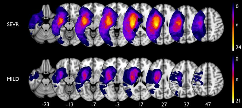
Numerous longitudinal imaging studies have aimed to explore recovery of stroke patients and relate it to time-dependent alterations of structural and functional brain networks. However, the majority of studies though have included only moderately to mildly affected patients. Whether our present concepts will also be present in more severely affected patients remains unclear. The present longitudinal and multimodal study seeks to close this gap of knowledge. We will include acute first-ever stroke patients and conduct clinical and imaging-based follow-up during multiple time points during the first year of recovery. Brain imaging will consist of structural and functional MRI. Clinical testing will consist of various score to assess different aspects of motor functioning and recovery.
This project is funded by the Else Kröner-Fresenius-Stiftung.
Related Publications:
Frontzkowski L, Fehring F, Frey BM, Wrobel PP, Reibelt A, Higgen F, Wolf S, Backhaus W, Braass H, Koch PJ, Choe CU, Bonstrup M, Cheng B, Thomalla G, Gerloff C, Quandt F, Schulz R. Frontoparietal Structural Network Disconnections Correlate With Outcome After a Severe Stroke. Hum Brain Mapp. 2024;45:e70060. doi: 10.1002/hbm.70060.
Nemati PR, Backhaus W, Feldheim J, Bonstrup M, Cheng B, Thomalla G, Gerloff C, Schulz R. Brain network topology early after stroke relates to recovery. Brain Commun. 2022;4:fcac049. doi: 10.1093/braincomms/fcac049.
Backhaus W, Braass H, Higgen FL, Gerloff C, Schulz R. Early parietofrontal network upregulation relates to future persistent deficits after severe stroke-a prospective cohort study. Brain Commun. 2021;3:fcab097. doi: 10.1093/braincomms/fcab097.
Diffusion-MRI to assess cortical microstructure

Characterizing cortical plasticity becomes increasingly important for identifying compensatory mechanisms and structural reserve in the ageing population. While cortical thickness (CT) largely contributed to systems neuroscience, it incompletely informs about the underlying neuroplastic pathophysiology. In turn, microstructural characteristics may correspond to pathological mechanisms in a more sensitive way. For instance, fractional anisotropy (FA), a diffusion tensor imaging (DTI) measure, is inversely related to cortical histologic complexity. Axial diffusivity (AD) and radial diffusivity (RD) are assumed to be linked to the density of structures oriented perpendicular and parallel to the cortical surface, respectively. We use 3T DTI data to assess cortical microstructural properties to better understand, e.g., patterns of hemispheric specialization, ageing, the impact of cardiovascular risk factors on brain health and trajectories of clinical cohorts with various neurological diseases.
Related Publications:
Wróbel PP, Guder S, Feldheim JF, Graterol Pérez JA, Frey BM, Choe C-u, Bönstrup M, Cheng B, Rathi Y, Pasternak O, Thomalla G, Gerloff C, Shenton ME, Schulz R. Assessing the cortical microstructure in contralesional sensorimotor areas after stroke. Brain Commun. 2024;6. doi: 10.1093/braincomms/fcae115.
Wrobel PP, Braass H, Frey BM, Bonstrup M, Guder S, Frontzkowski LK, Feldheim JF, Cheng B, Rathi Y, Pasternak O, Thomalla G, Koerte IK, Shenton ME, Gerloff C, Quandt F, Higgen FL*, Schulz R*. Cortical microstructure and hemispheric specialization-A diffusion-imaging analysis in younger and older adults. Eur J Neurosci. 2024;60:5718-5730. doi: 10.1111/ejn.16518.
Simultaneous cerebral and spinal cord functional MRI

Functional MRI is a powerful tool to study stroke-related changes in brain activation and information flow through multisite distributed brain networks. However, until recently, due to technical limitations, the field of view of functional MRI in stroke recovery research was limited to the brain. The complete corticospinal brain network, spanning from the motor cortices in the brain to spinal neuronal assemblies, has escaped from systematic functional analyses. However, recently, the interest in the corticospinal system and the spinal cord after stroke experienced a remarkable renaissance. Recent advancements in functional MRI of the spinal cord and simultaneous corticospinal functional MRI are opening a new window to investigate functional corticospinal networks in humans with high spatial resolution. In a first exploratory proof-of-concept study, we could link ventral spinal activation during simple hand movements not only to M1 activation but also to ventral premotor cortex activation. Currently, we are applying simultaneous corticospinal functional MRI in stroke patients to explore stroke-related changes in activation and coupling between cerebral and spinal neuronal assemblies.
Related Publications:
Braass H, Feldheim J, Chu Y, Tinnermann A, Finsterbusch J, Buchel C, Schulz R*, Gerloff C*. Association between activity in the ventral premotor cortex and spinal cord activation during force generation-A combined cortico-spinal fMRI study. Hum Brain Mapp. 2023. doi: 10.1002/hbm.26523.
Oscillatory network modulation to enhance motor skill acquisition
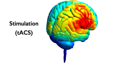
Transcranial alternating current stimulation (tACS) has been shown to entrain neurons in recent macaque studies and to modulate oscillatory connectivity as well as human behaviour. Based on knowledge gained from our studies on the neuronal coding of motor control we aim to influence the motor network to enhance motor performance in stroke patients. Based on the assumption that theta-gamma phase amplitude coupling is a key mechanism for motor skill acquisition, we currently investigate the impact of tACS on motor skill acquisition in healthy participants and stroke patients. The studies are in close collaboration with Dr. Bettina Schwab.
OSF: https://doi.org/10.17605/OSF.IO/MQWT5, Quandt & Schwab
ClinicalTrials.gov NCT05576129, Quandt & Schwab.
Oscillatory modulation of the parietofrontal network to enhance motor performance after stroke

In this project we will apply bi-focal high-definition tACS covering posterior-parietal brain regions and the primary motor cortex of the ipsilesional hemisphere to enhance parietofrontal network connectivity and improve motor functions in chronic stroke patients. MRI and EEG analyses will be used to understand inter-subject variability in behavioural tACS-responses.
This project is funded by the Damp-Stiftung.
Related publication:
Reibelt A, Quandt F, Schulz R. Posterior parietal cortical areas and recovery after motor stroke: a scoping review. Brain Commun. 2023;5:fcad250. doi: 10.1093/braincomms/fcad250
ProbeAIP – Virtual lesioning of the anterior intraparietal sulcus by TMS
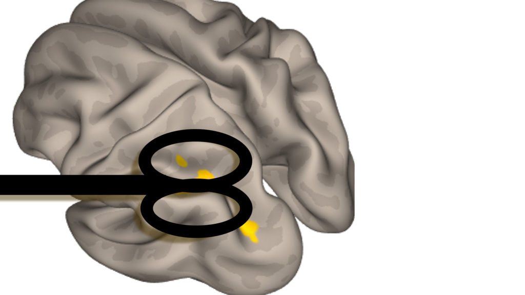
The is increasing evidence that the ipsilesional anterior intraparietal sulcus (aIPS) might be critically involved in residual motor functioning and recovery processes after stroke. However, for previous imaging-based studies it remained an open question to what extent the aIPS might directly act onto network dynamics and clinical aspects or whether it may exert its influence indirectly via other brain networks or hubs, i.e. the causal influence of aIPS fin brain dynamics and motor behavior has yet to be clarified. This study investigates chronic stroke patients and uses transcranial magnetic stimulation (TMS) to perturbate aIPS during simple motor tasks. EEG and behavioral testing will be used to quantify the effects of this virtual lesioning. Structural and functional MRI data will be acquired to adress associations between brain network architecture and TMS effects.
This project was funded by the Werner-Otto-Stiftung.
MARK-NIBS – Network Properties as BioMARKers for Non-Invasive Brain Stimulation After Stroke
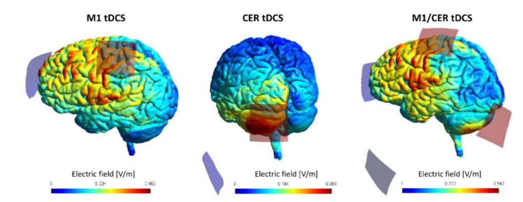
Various studies have aimed to explore the potential of non-invasive brain stimulation techniques such as transcranial direct current stimulation (tDCS) to promote motor recovery after stroke. After promising results from early proof-of-concept studies, particularly for the stimulation of the primary motor cortex (M1), it has become evident that the translation from scientific to clinical application is challenging. Aiming to uncover alternative stimulation targets, the cortico-cerebellar network and cerebellar brain stimulation have gained an increasing interest in the field of neurorehabilitation. However, large inter-study and inter-subject variability in behavioural responses to tDCS indicated that a one-size-fits-all approach might not lead to sufficient effect sizes in clinical populations. As structural and functional brain imaging has significantly evolved to powerful tools to assess distinct neuronal networks, such as the cortico-cerebellar network, in individual stroke patients and to infer structure-function-behaviour-relationships, the question arises whether such information might serve as imaging biomarkers to inform about the treatment responsiveness to non-invasive brain stimulation. This study will evaluate the potential of cortico-cerebellar network properties in a group of chronic stroke patients and healthy participants to explain inter-subject variability in responsiveness to two brain stimulation approaches targeting the cortico-spinal and cortico-cerebellar network: 1) cortical M1 tDCS, 2) combined M1 and cerebellar tDCS. Participants are examined clinically and by structural and functional MRI. Structural MRI is used to primarily reconstruct cortico-spinal and cortico-cerebellar motor tracts. Tract-related diffusion-based parameters are used to infer microstructural network integrity. Resting-state MRI data are acquired to assess functional network connectivity. The behavioural impact of the tDCS is evaluated during a multi-session structured motor training paradigm over seven days. Statistics integerate clinical treatment responses with structural and functional network infromation to better understand tDCS responses on a single-subject level.
ClinicalTrials.gov NCT05560724
Related publications:
Guder S, Frey BM, Backhaus W, Braass H, Timmermann JE, Gerloff C, Schulz R. The Influence of Cortico-Cerebellar Structural Connectivity on Cortical Excitability in Chronic Stroke. Cereb Cortex. 2020;30:1330-1344. doi: 10.1093/cercor/bhz169.
Schulz R*, Frey BM*, Koch P, Zimerman M, Bonstrup M, Feldheim J, Timmermann JE, Schon G, Cheng B, Thomalla G, Gerloff C, Hummel FC. Cortico-Cerebellar Structural Connectivity Is Related to Residual Motor Output in Chronic Stroke. Cereb Cortex. 2017;27:635-645. doi: 10.1093/cercor/bhv251.
StimAIP – Non-invasive brain stimulation to the posteroparietal cortex to enhance residual motor functions in chronic stroke
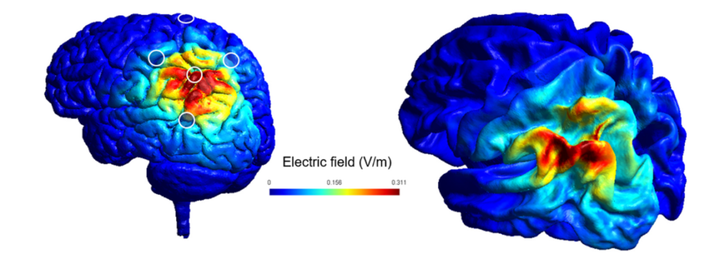
Previous studies have aimed to explore the potential of non-invasive brain stimulation techniques such as transcranial direct current stimulation (tDCS) to promote motor recovery after stroke. After promising results from early proof-of-concept studies, particularly for the stimulation of the primary motor cortex, it has become evident that the translation from scientific to clinical application is challenging. Aiming to uncover alternative innovative stimulation targets, recent reports suggest that the anterior intraparietal sulcus (AIPS) might be crucially involved in aspects of residual motor functioning and also recovery processes after stroke. In the present project, chronic stroke patients will undergo high-definition activating tDCS of the ipsilesional AIPS to promote motor capabilities. The participants will be examined by means of clinical testing. Transcranial magnetic stimulation will be used to test the impact of AIPS tDCS to network excitability. Structural and functional MRI will be used to assess the relationship between network structure and function and the responsiveness to AIPS stimulation. As an overarching goal, this study might provide data towards studies investigating patient stratification protocols providing individual treatment strategies to individual patients.
This project is funded by the Else Kröner-Fresenius-Stiftung.
Related publications:
Reibelt A, Quandt F, Schulz R. Posterior parietal cortical areas and recovery after motor stroke: a scoping review. Brain Commun. 2023;5:fcad250. doi: 10.1093/braincomms/fcad250
Outcome prediction in large clinical stroke cohorts

We employ advanced machine-learning techniques to predict outcome after stroke, focusing on the impact of stroke treatment on functional outcome. To achieve our goal, we analyse multicenter databases such as the German Stroke Registry and the VISTA database – endovascular. This project is in collaboration with the CSI-Lab as well as the Tiedt-Lab (Steffen Tiedt, LMU Munich).
Related Publications:
Burbano, V. G., Wolfer, T. A., Vlegels, N., Quandt, F., Zimmermann, H., Wischmann, J., Kellert, L., Liebig, T., Dimitriadis, K., Saver, J. L., Tiedt, S., & investigators, G. S. R. (2023). Association of the time of day of EVT with clinical outcomes and benefit from successful recanalization after stroke. Ann Clin Transl Neurol. https://doi.org/10.1002/acn3.51877
Quandt, F., Flottmann, F., Madai, V. I., Alegiani, A., Kupper, C., Kellert, L., Hilbert, A., Frey, D., Liebig, T., Fiehler, J., Goyal, M., Saver, J. L., Gerloff, C., Thomalla, G., Tiedt, S., investigators, G. S. R., & the, V.-E. C. (2023). Machine Learning-Based Identification of Target Groups for Thrombectomy in Acute Stroke. Transl Stroke Res, 14(3), 311-321. https://doi.org/10.1007/s12975-022-01040-5
Quandt, F., Meissner, N., Wolfer, T. A., Flottmann, F., Deb-Chatterji, M., Kellert, L., Fiehler, J., Goyal, M., Saver, J. L., Gerloff, C., Thomalla, G., & Tiedt, S. (2023). RCT versus real-world cohorts: Differences in patient characteristics drive associations with outcome after EVT. Eur Stroke J, 8(1), 231-240. https://doi.org/10.1177/23969873221142642
IMPROVE – Interdisciplinary Platform for Rehabilitation Research and Innovative Care of Stroke Patients
IMPROVE is an Interdisciplinary Platform for Rehabilitation Research and Innovative Care of Stroke Patients. The aim of this collaborative platform is to foster neurorehabilitation research and to transfer evidence-based innovative neurorehabilitation into clinical practice. As a first step, an observational, longitudinal, multicenter study of functional stroke recovery after patients’ discharge from inpatient rehabilitation is performed to better understand the dynamics and underlying mechanisms of recovery from stroke.
IMRPOVE is a cooperation between the University Medical Center Hamburg-Eppendorf (UKE), five Neurological Rehabilitation Centers in Northern Germany (RehaCentrum Hamburg, MEDICLIN Klinikum Soltau, Klinikum Bad Bramstedt, VAMED Klinik Geesthacht and VAMED Rehaklinik Damp), and the Deutsche Rentenversicherung Nord (DRV Nord).
ClinicalTrials.gov NCT04119479