Cross-frequency coupling related to motor skill acquisition in stroke patients
The project aims to study the neurophysiological basis, in particular oscillatory changes, of motor skill learning in stroke patients with hand motor deficits. In a multimodal approach, we explore signatures of neural activity in stroke patients related to motor skill acquisition with a special interest in the role of cross-frequency coupling. We combine MEG and behavioral recordings of a motor learning paradigm with structural MRI data. Our results are thought to lay the grounds to enhance mechanistically informed and individualized treatment strategies via non-invasive brain stimulation to promote motor recovery in stroke patients.
This is project is conducted in collaboration with the Department of Neurophysiology and Pathophysiology (Dr. Bettina Schwab) and funded by internal funds of the UKE.
Crossmodal learning
Multisensory integration in the aging brain — mechanisms and facilitation

The overall goal of the project is to examine the neural dynamics of multisensory integration in healthy older adults. We analyze neural signals associated with visuo-tactile integration and learning using EEG and MEG. Furthermore, we employ non-invasive brain stimulation to modulate related brain areas in attempt to enhance performance and aim to build computational models accounting for age-related differences. Our investigations will focus specifically on oscillatory neural activity and large-scale dynamic coupling between brain regions.
This project ist part of the Transregional Collaborative Research Centre – Crossmodal Learning: Adaptivity, Prediction and Interaction (TRR 169) and funded by the German Research Foundation (DFG) and the National Natural Science Foundation of China (NSFC).
Crossmodal learning in health and neurological disease: neurocomputational representation and therapeutic application

This project constitutes the follow-up to the project “Multisensory integration in the aging brain – mechanisms and facilitation”. Here, we aim at providing deeper neurocomputational insight into the alterations of crossmodal integration in healthy older adults and extending our work to neurological diseases (i.e. stroke and Parkinson’s disease). We determine the interrelation between the structural and functional changes in multisensoryl brain networks by means of simultaneously recorded EEG-fMRI activation data in healthy older adults and neurological patients. Moreover, we use enhanced sensory feedback and virtual reality (VR) enhanced training to modulate and, ideally, improve performance in crossmodal integration tasks.
This project ist part of the Transregional Collaborative Research Centre – Crossmodal Learning: Adaptivity, Prediction and Interaction (TRR 169) and funded by the German Research Foundation (DFG) and the National Natural Science Foundation of China (NSFC).
KETCH – Encoding of kinematics and effector choice in reorganized brain networks after stroke
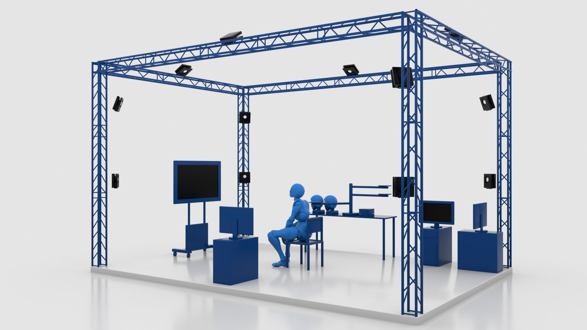
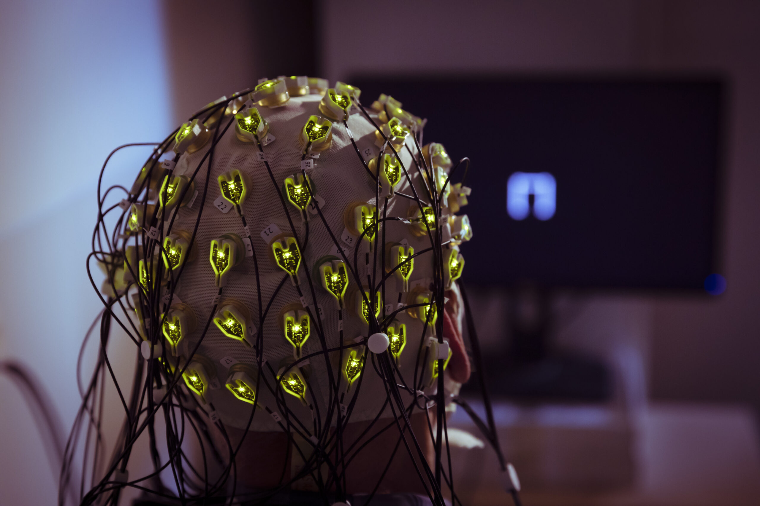
This project has the principal goal of advancing our understanding of how neuronal networks in the human brain reorganize after a stroke and how this affects complex motor behavior relevant to everyday life. We try to gain new insights into motor-related cognitive processes, and to expand our network models through a technically innovative analysis of the spinal cord’s activity. By using an advanced system of infrared 3D cameras while measuring EEG signals we are able to study kinematic parameters, such as “smoothness” and “directness” of the movements, and to correlate them with the brain activity of the participants. Additionally, we are able to study the activation in predefined cortico-spinal networks with special fMRI-sequences. In addition to gaining fundamental scientific knowledge, we also study the neuroplasticity-enhancing effects of targeted neuromodulation of neural networks.
This project is part of the Collaborative Research Center 936 – Multi-Site Communication in the Brain (CRC 936/SFB 936) and funded by the Deutsche Forschungsgesellschaft (DFG). DFG Project number, 178316478
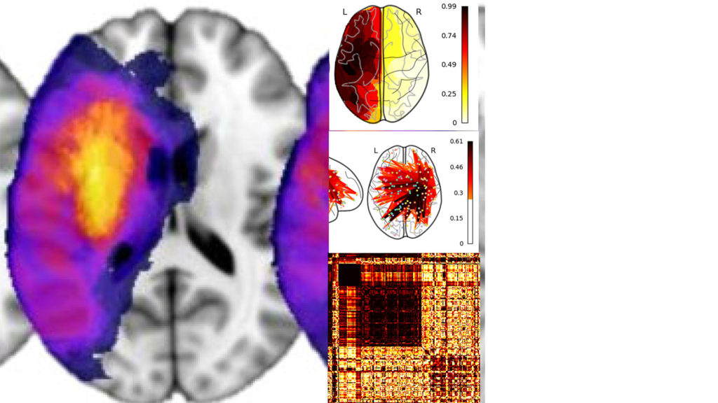
MARK-NIBS – Network Properties as BioMARKers for Non-Invasive Brain Stimulation (NIBS) After Stroke
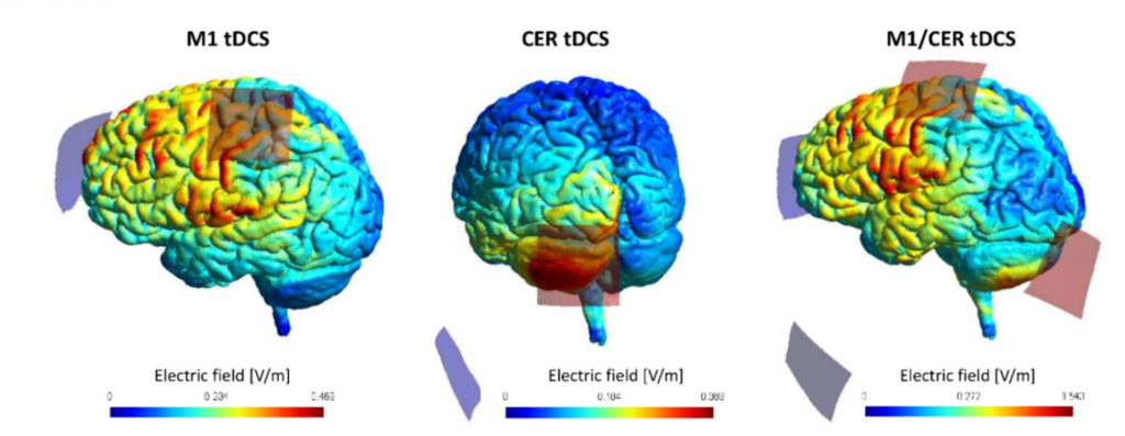
Various studies have aimed to explore the potential of non-invasive brain stimulation techniques such as transcranial direct current stimulation (tDCS) to promote motor recovery after stroke. After promising results from early proof-of-concept studies, particularly for the stimulation of the primary motor cortex (M1), it has become evident that the translation from scientific to clinical application is challenging. Aiming to uncover alternative stimulation targets, the cortico-cerebellar network and cerebellar brain stimulation have gained an increasing interest in the field of neurorehabilitation. However, large inter-study and inter-subject variability in behavioural responses to tDCS indicated that a one-size-fits-all approach might not lead to sufficient effect sizes in clinical populations. As structural and functional brain imaging has significantly evolved to powerful tools to assess distinct neuronal networks, such as the cortico-cerebellar network, in individual stroke patients and to infer structure-function-behaviour-relationships, the question arises whether such information might serve as imaging biomarkers to inform about the treatment responsiveness to non-invasive brain stimulation. The present study will evaluate the potential of cortico-cerebellar network properties in a group of chronic stroke patients and healthy participants to explain inter-subject variability in responsiveness to two brain stimulation approaches targeting the cortico-spinal and cortico-cerebellar network: 1) cortical M1 tDCS, 2) combined M1 and cerebellar tDCS. Participants will be examined clinically and by structural and functional MRI. Structural MRI will be used to primarily reconstruct cortico-spinal and cortico-cerebellar motor tracts. Tract-related diffusion-based parameters will be used to infer microstructural network integrity. Resting-state MRI will be acquired to assess functional network connectivity. The behavioural impact of the tDCS will be evaluated during a multi-session structured motor training paradigm over seven days. Statistics will integerate clinical treatment responses with structural and functional network infromation to better understand tDCS responses on a single-subject level.
ClinicalTrials.gov NCT05560724
ProbeAIP – Virtual lesioning of the aterior intraparietal sulcus by TMS to assess its influence for network dynamics and motor function after stroke
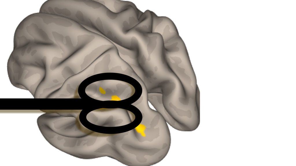
The is increasing evidence that the ipsilesional anterior intraparietal sulcus (aIPS) might be critically involved in residual motor functioning and recovery processes after stroke. However, for previous imaging-based studies it remained an open question to what extent the aIPS might directly act onto network dynamics and clinical aspects or whether it may exert its influence indirectly via other brain networks or hubs, i.e. the causal influence of aIPS fin brain dynamics and motor behavior has yet to be clarified. The present study will investigate chronic stroke patients and use transcranial magnetic stimulation (TMS) to perturbate aIPS during simple motor tasks. EEG and behavioral testing will be used to quantify the effects of this virtual lesioning. Structural and functional MRI will be applied to assess the association between brain network architecture and the TMS effects.
This project is funded by the Werner-Otto-Stiftung.
SEVERE – Longitudinal multimodal assessment of recovery of severely impaired stroke survivors
Numerous longitudinal imaging studies have aimed to explore recovery of stroke patients and relate it to time-dependent alterations of structural and functional brain networks. However, the majority of studies though have included only moderately to mildly affected patients. Whether our present concepts will also be present in more severely affected patients remains unclear. The present longitudinal and multimodal study seeks to close this gap of knowledge. We will include acute first-ever stroke patients and conduct clinical and imaging-based follow-up during multiple time points during the first year of recovery. Brain imaging will consist of structural and functional MRI. Clinical testing will consist of various score to assess different aspects of motor functioning and recovery. If visits at our hospital will not be possible, we will visit the patients at home.
This project is funded by the Else Kröner-Fresenius-Stiftung.
StimAIP – Non-invasive brain stimulation to the posteroparietal cortex to enhance residual motor functions in chronic stroke

Previous studies have aimed to explore the potential of non-invasive brain stimulation techniques such as transcranial direct current stimulation (tDCS) to promote motor recovery after stroke. After promising results from early proof-of-concept studies, particularly for the stimulation of the primary motor cortex, it has become evident that the translation from scientific to clinical application is challenging. Aiming to uncover alternative innovative stimulation targets, recent reports suggest that the anterior intraparietal sulcus (AIPS) might be crucially involved in aspects of residual motor functioning and also recovery processes after stroke. In the present project, chronic stroke patients will undergo high-definition activating tDCS of the ipsilesional AIPS to promote motor capabilities. The participants will be examined by means of clinical testing. Transcranial magnetic stimulation will be used to test the impact of AIPS tDCS to network excitability. Structural and functional MRI will be used to assess the relationship between network structure and function and the responsiveness to AIPS stimulation. As an overarching goal, this study might provide data towards studies investigating patient stratification protocols providing individual treatment strategies to individual patients.
This project is funded by the Else Kröner-Fresenius-Stiftung.